SDS-PAGE
Any ion or charged group will migrate when placed in an electric field. Since proteins carry a net charge at any pH other than their isoelectric point, they too will migrate and their rate of migration will depend upon the charge density (the ratio of charge to mass) of the proteins concerned; the higher the ratio of charge to mass the faster the molecule will migrate. In zone electrophoresis, the mixture of molecules to be separated is placed as a narrow zone or band at a suitable distance from the electrodes such that, during electrophoresis, proteins of different mobilities travel as discrete zones which gradually separate from each other as electrophoresis proceeds. In practice, zone electrophoresis of proteins is rarely carried out in free solution but instead is performed in a solution stabilized within a supporting medium. Many supporting media are in current use but easily the most popular for electrophoresis of proteins is polyacrylamide gel.
Polyacrylamide gel results from the polymerization of acrylamide monomer into long chains and the cross-linking of these by bifunctional compounds such as N,N'-methylene bisacrylamide (usually abbreviated to bisacrylamide). This reaction creates a porous matrix in which the pore size is the same order of size as protein molecules. Thus, during electrophoresis in polyacrylamide gel, the proteins are subject to a molecular sieving effect which slows down the migration of larger proteins relative to smaller proteins. Separation of native proteins by polyacrylamide gel electrophoresis (PAGE) in non-dissociating buffers (e.g. Tris/HCI) is therefore dependent on both charge density and size.
The extent of molecular sieving during PAGE depends on how closely the gel pore size approximates the size of the migrating proteins. The effective pore size of polyacrylamide gel varies both with the total concentration of acrylamide in the gel mixture (pore size decreasing as the acrylamide concentration increases) and with the proportion of the bisacrylamide cross-linker used. In practice, the proportion of cross-linker is held constant and the effective pore size is varied by altering the concentration of acrylamide monomer. Most protein separations are carried out using gels ranging from 5 to 15% acrylamide. The lower percentage gels have the largest pore sizes and so, for zone electrophoresis, are used to separate larger proteins whereas the acrylamide concentration is increased to resolve smaller proteins.
Originally, zone electrophoresis of proteins in polyacrylamide made use of cylindrical rod gels in glass tubes but nowadays flat slab gels, 0.75 - 1.5 mm thick, are usually preferred instead. One of the main advantages of slab gels is that many protein samples, including molecular weight marker proteins (see below), can be electrophoresed under identical conditions in a single gel so that the band patterns produced are directly comparable. In contrast, due to minor differences in polymerisation efficiency, gel length, gel diameter, etc., rod gels even of the same protein sample are rarely identical. Although slab gels are now used almost to the exclusion of rod gels for simple electrophoretic separations, rod gels are preferred for certain situations, especially to carry out the first-dimensional step of two-dimensional gel electrophoresis.
Although PAGE of native proteins is useful in a number of situations, especially where one wishes to detect a particular separated protein by its biological activity, the vast majority of studies involving PAGE use conditions designed to dissociate all proteins in the sample into their individual polypeptide subunits. The dissociation agent most commonly used is the ionic detergent, sodium dodecyl sulphate (SDS). SDS-PAGE of proteins allows rapid analysis of the composition of protein mixtures and so is extremely useful for monitoring the purity of protein samples during purification protocols. For certain applications, it can even be used as a small-scale preparative procedure.
For analysis by SDS-PAGE, the protein mixture is denatured by heating at 100
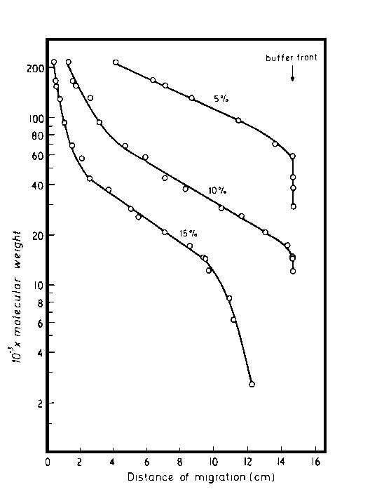
°C for 2 - 5 minutes in the presence of excess SDS and thiol reagent such as 2-mercaptoethanol (to cleave disulphide bonds). Under these conditions, most polypeptides bind SDS in a constant weight ratio (1.4g SDS per gram of polypeptide). Each sample is then loaded onto a separate rod gel or into a separate well of the slab gel and electrophoresis is carried out with SDS present both in the gel and in the electrode buffer. For reasons discussed below, marker proteins of known molecular weight are also dissociated in an identical manner and loaded into other wells. After electrophoresis, the positions of the separated protein bands are revealed by staining, usually with Coomassie Blue or a silver stain.During SDS-PAGE, the intrinsic charges of the polypeptides are insignificant compared with the negative charges provided by the bound detergent, so that the SDS-polypeptide complexes have essentially identical charge densities and migrate in polyacrylamide gels of the correct porosity strictly according to polypeptide size and not charge. Since the distance migrated by a polypeptide is directly proportional to its size, the protein of interest can be identified in the gel profile if the molecular weight of its constituent polypeptides is known or, conversely, the molecular weight of individual polypeptides can be determined. To estimate the molecular weight of a sample polypeptide requires the investigator to construct a standard curve of log(polypeptide molecular weight) versus distance migrated for each of the standard molecular weight marker polypeptides run on the gel. This yields a straight line relationship. Knowing the distance migrated by the sample polypeptide, one can then read off its molecular weight from the curve.
Calibration curves of log
10(polypeptide molecular weight) versus distance of migration during SDS-PAGE in polyacrylamide slab gels. The polyacrylamide gels used were uniform concentration 5%, 10% or 15%. [Reproduced from Hames,B.D. (1981) In Gel Electrophoresis of Proteins A Practical Approach. Hames,B.D. and Rickwood,D. (eds), IRL Press Ltd, Oxford.]
Note, however, that for any one gel concentration the relationship between log(molecular weight) and distance migrated (or relative mobility) is linear over only a limited range of molecular weight, so that the gel concentration chosen for the step must be one which will yield a linear relationship in the desired molecular weight range. This problem, which occurs with gels of uniform acrylamide concentration, can be overcome to a large extent by using concentration gradient gels where the acrylamide concentration increases continuously (and hence pore size decreases continuously) with increasing migration distance. Despite this advantage, it is important to note that gradient gels cannot match the resolution of two protein components obtainable with a gel of uniform optimal polyacrylamide concentration.
The maximum capacity of each gel track of a slab gel required to obtain good resolution is such that only 1 - 10 micrograms of each polypeptide or 50 - 100 micrograms of a complex mixture can be loaded. Even using a complete slab gel to load a single protein sample can barely yield milligram quantities of a pure polypeptide component. Therefore, when applied during a protein purification scheme, SDS-PAGE is used mainly to monitor the progress of the purification, i.e. to assess the composition and purity of collected fractions. The exception, of course, is when only small amounts of the desired protein need to be purified. For example, modern methods of protein sequencing can yield very substantial data using sub-microgram quantities of protein. Furthermore these protein sequencing techniques can operate effectively using SDS-polypeptide cornplexes. Therefore it may well be sufficient to electrophorese a partially purified protein sample on an SDS-polyacrylamide gel and then, after electrophoresis, to recover the separated polypeptide of interest by elution from the gel. In cases such as this, SDS-PAGE is used as a small-scale preparative method. However, this aspect is not simulated in this program.